- APPLY FOR SLOT
- Internal Users
- External Users
- SLOT BOOKING STATUS
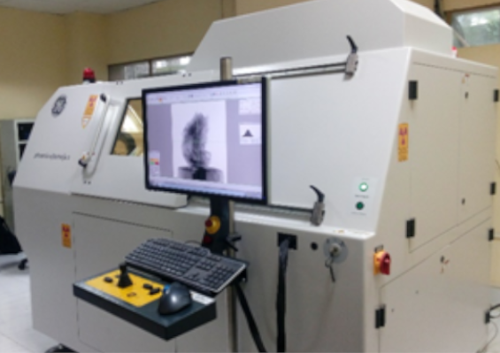
Non-Destructive Imaging Laboratory (X-Ray Micro-CT)
Phone : +91-3222-282306
Location : OB / GF / 4, CRF
Facilitator :
Prof. Santanu Dhara, Medical Science and Technology
Email: sdhara@smst.iitkgp.ac.in, Contact:+91-3222-282306
For Internal Users - Click Here to apply for Slot
For External Users - Click Here to apply for Slot
Objectives
Micro and Nano CT scanning device creates 2D slices of an object based on material density measured by X-ray transmission. Various type of samples of different attributes and structures ranging from geology, space technology, materials manufacturing, biomedical are analyzed in micro nano computed tomography. In domain of electronic components, micro devices, composites, porous metallic/ceramic, polymers and rubber components, biomedical samples, food, micro fossils are also analyzed. The visualization is generated for depth information of porosity analysis. The other parameters such as analysis microstructure, pore size distribution, fiber volume analysis and textured analysis are also done by using analyzing software. Besides these, the image reconstruction and analysis of exact size and shape of any 3D objects would be helpful via reverse engineering tool. Non-destructive imaging, FE analysis and development of implants and devices, automotive engine, mechanical device manufacturing can also be facilitated using these tool.
People
Prof. Santanu Dhara
Facilitator
Medical Science and Technology
sdhara@smst.iitkgp.ac.in
+91-3222-282306
Equipment Details
Utility and Working Principal
In micro CT (computed tomography) a micro-CT image is generated by rotating the sample in very small steps around a single axis of rotation while taking a series of 2D radiographs. The 3D image can reconstructed using reconstruction algorithms, such as filtered back projection algorithm, and saved as volume file. The voltage and current are also selected for x ray generation depending on the density of the material. After selection of following parameters detector calibration step is done to calibrate the detector. In order to do this step the sample is moved out from the line of X-ray focus. After calibration the sample moves automatically to its original saved position. Once the sample moves to original position the scan starts.
The acquisition file is obtained and further processed for analysis purpose. The file is exported to analysis software for volume rendering. Surface determination is done by selecting the background and material in the 2D image. After calculating, 3D view of the sample is obtained. To characterize the material and pores in the material, porosity analysis module is used. The pores are identified by respective color codes with percentage of volume.
Sample Details
Sample preparation
- No sample preparation is required.
Precautions:
- Radioactive elements should be avoided.
- Samples having high density should be avoided.
- Operator should follow proper laboratory procedures in handling the equipment.

CRF IIT Kharagpur
To offer research oriented support to academic and research institutes, industries (R&D) etc.
Facilitating institute and industry collaboration by providing crucial services for sponsored research and consulting.
Create a program to host short-term seminars, workshops, and training sessions on the use of analytical methodologies for students, researchers, instructors, and industry professionals.
Contact Us
EXPLORE CRF
- © All Rights Reserved, CRF IIT Kharagpur
List of Laboratories
- 1. Micro and Nano Scratch Testing Lab
- 2. Universal Testing Machine (UTM)
- 3. Vickers Micro-Hardness Lab
- 4. HR TEM Laboratory
- 5. 2D-X-Ray Diffraction Lab
- 6. X-ray Photoelectron Spectroscopy Laboratory
- 7. Analytical Ultracentrifuge Lab
- 8. ZEISS Scanning Electron Microscope Laboratory (EVO 60)
- 9. Vibrating Sample Magnetometer Laboratory
- 10. Thermal Analysis (DSC, DTA, TGA, TMA) Laboratory
- 11. TEM Sample Preparation Laboratory
- 12. SQUID Laboratory
- 13. Single Crystal X-ray Diffractometer Laboratory
- 14. Small Angle X-ray Scattering Laboratory
- 15. Raman Spectrometer Laboratory
- 16. Pulsed Electron Spin Paramagnetic Resonance Laboratory
- 17. Optical Microscopy and Mechanical Testing Laboratory
- 18. Nano - Indentation & Nano - Tribology Laboratory
- 19. Non-Destructive Imaging Laboratory (X-Ray Micro-CT)
- 20. INSTRON Laboratory
- 21. High Resolution - XRD Laboratory
- 22. Micro Raman Spectrometer Laboratory
- 23. Hall Effect Laboratory
- 24. Fourier Transform-Infrared Spectrometer Laboratory
- 25. Cross/Dual beam FIB-FEG microscopy Laboratory(FIB Lab)
- 26. Field Emission Gun-Scanning Electron Microscope Laboratory (Zeiss Supra 40)
- 27. Field Emission Gun-Scanning Electron Microscope Laboratory (Merlin)
- 28. FEG-High Resolution-Cryo-Analytical TEM Laboratory
- 29. Scanning Auger Nanoprobe Laboratory
- 30. Analytical Transmission Electron Microscope Laboratory
- 31. Atomic Force Microscope Laboratory
- 32. 3D - Optical Surface Profilometer Laboratory
- 33. X-ray Protein Crystallography Laboratory
- 34. Real Time PCR Laboratory
- 35. Nuclear Magnetic Resonance Laboratory (600 MHz)
- 36. Matrix Assisted Laser Desorption Ionization Laboratory (MALDI-ToF MS)
- 37. Liquid Chromatography - MS / MS Laboratory
- 38. Isothermal Titration Calorimetry Laboratory
- 39. High Resolution - Mass Spectrometry Laboratory (HRMS)
- 40. High Performance Liquid Chromatography Lab (HPLC)
- 41. Fluorescence Activated Cell Sorter Lab (FACS)
- 42. DNA Sequencer Laboratory
- 43. Circular Dichroism Spectropolarimetry Laboratory
- 44. 2D - GEL Electrophoresis Laboratory